
Anti-Goat IgG (H+L) – Gold Conjugate
3rd Nov 2026
GOLD CONJUGATED ANTIBODIES
Anti-Goat IgG (H+L) – Gold Conjugate
Rabbit Polyclonal Antibody | Gold Nanoparticle Conjugate | Secondary Detection
Affinity-purified antibody coupled to gold nanoparticles
Product Information
Product: Anti-Goat IgG (H+L) – Gold Conjugate
Antibody Host: Rabbit
Target: Goat IgG (Heavy and Light Chains)
Conjugate: Gold Nanoparticles
Clonality: Polyclonal
Purification: Affinity Isolated
Concentration: 0.15 mg/mL (@ OD=3); 0.5 mg/mL (@ OD=10)
Working Dilution: 1:10–1:100
Storage: 2–8°C – Do Not Freeze
Application: Research Use Only
Product Description
Anti-Goat IgG (H+L) Gold Conjugate is an affinity-isolated rabbit polyclonal antibody coupled to gold nanoparticles. It is designed for the secondary detection of samples containing goat-derived antibodies in a variety of immunological and microscopy applications.
The gold nanoparticle conjugate provides a sensitive and stable labeling system for detecting goat IgG in experimental samples. Its applications include lateral flow assays, immunoblotting, light microscopy and electron microscopy.
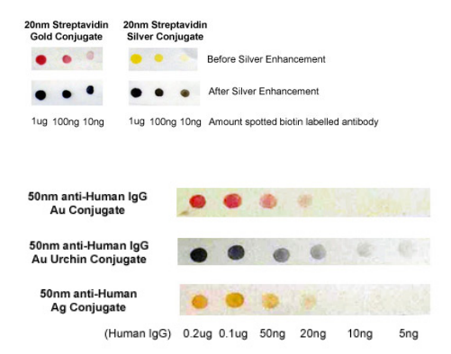
Gold conjugates can also be used in combination with silver enhancement reagents to increase signal visibility and detection sensitivity. This makes the conjugate useful for immunogold labeling procedures and membrane-based detection techniques.
The product can be used independently or alongside silver enhancer kits for membrane and microscopy applications.
Key Features
- Affinity-isolated rabbit anti-goat IgG antibody.
- Recognizes goat IgG heavy and light chains (H+L).
- Conjugated to gold nanoparticles.
- Polyclonal antibody formulation.
- Suitable for secondary antibody detection.
- Compatible with silver enhancement procedures.
- Suitable for membrane-based and microscopy applications.
- Working dilution range: 1:10–1:100, depending on the application.
Applications
- Lateral Flow Assays: Gold nanoparticle-based secondary antibody detection.
- Immunoblotting: Detection of goat primary antibodies on membranes.
- Light Microscopy: Visualization of immunogold-labeled targets.
- Electron Microscopy: Gold nanoparticle labeling for ultrastructural studies.
- Dot Blot: Detection of immobilized proteins using immunogold labeling.
- Silver Enhancement: Signal amplification for improved visualization.
Technical Specifications
Product Name: Anti-Goat IgG (H+L) – Gold Conjugate
Antibody Host: Rabbit
Antibody Specificity: Goat IgG (H+L)
Conjugated Antibody: Rabbit affinity-purified anti-goat IgG (H+L)
Clonality: Polyclonal
Conjugate: Gold nanoparticles
Concentration: 0.15 mg/mL (@ OD=3)
Concentration: 0.5 mg/mL (@ OD=10)
Working Dilution: 1:10–1:100
Storage Buffer: 20 mM Tris (pH 8.0), 150 mM NaCl, 20% glycerol (v/v), 1% BSA
Storage and Stability
Store the product undiluted in its supplied storage buffer at 2–8°C.
Stability: Stable for at least 4 months when stored under the recommended conditions.
Important: DO NOT FREEZE. Freezing may affect the performance and stability of the gold conjugate.
Standard Immunogold Dot-Blot Protocol
The following procedure is adapted from Moeremans et al. (1984) and provides a general workflow for immunogold dot-blot detection.
- Spot 1 µL drops of serially diluted protein samples (1 µg–1 ng) in PBS supplemented with 0.5 µg/mL BSA onto a nitrocellulose or PVDF membrane.
- Allow the protein samples to dry and become immobilized on the membrane.
- Block the membrane for 30 minutes at room temperature using 1% (w/v) dry milk in 1X PBS.
- Incubate the membrane with the primary antibody for 2 hours at room temperature.
- Wash the membrane three times for 5 minutes each using the blocking solution.
- Incubate for 2 hours or longer with the gold-conjugated secondary antibody diluted 1:10 (OD=0.3) in blocking solution (0.2% Blocking Solution).
- Wash the membrane three times for 5 minutes each.
- Allow the membrane to dry and record the results.
- Optional: Perform silver enhancement to improve signal visibility and detection sensitivity.
Note: Working dilution and incubation conditions may require optimization depending on the sample, membrane and experimental application.
Related Products
Silver Enhancer Kit for Membranes
Silver Enhancer Kit for Microscopy
Product Safety and Handling
This product is intended for research and development use only. It is not intended for drug, household or other non-research uses.
Review the relevant Safety Data Sheet (SDS) before handling the product. Follow appropriate laboratory safety procedures and recommended storage conditions.
Scientific Reference
Moeremans M, et al. Journal of Immunological Methods. 1984; 74: 353.
This publication is cited as the methodological reference for the adapted immunogold dot-blot procedure.
Explore Gold-Conjugated Antibodies for Research
Discover antibody detection solutions for immunoblotting, microscopy and immunological research.
LOGIN FOR PRICE